On the right is a picture of two healthy teeth in cross section. These particular teeth happen to be a lower right first molar and second premolar. The teeth are supported in the bone by the “periodontal membrane” (see Healthy Gingiva ). The bone and the periodontal membrane are covered and protected by the “gingiva” or gums.
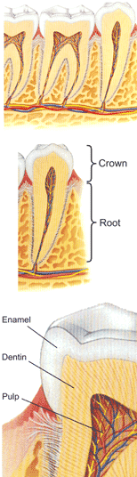
The tooth is divided into two major parts: The visible part of the tooth is the crown of the tooth. The part of the tooth hidden by the gingiva, and supported in the bone is the root. This premolar tooth has only one root, as do all of the front teeth. The molar teeth seen above are typical lower molars with two roots. The upper molars usually have three roots.
The tooth structure has three layers: The surface of the crown is covered by enamel. The enamel is the hardest substance in the human body. It is this hardness which allows us to chew food efficiently and to crunch through those harder foods like nuts and seeds. Most of the structure of the tooth is made up of dentin, which is not as hard as enamel, and is slightly flexible. In the centre of the tooth is the pulp chamber, which contains the dental pulp. The pulp is often referred to as the nerve, but it is, in fact, a mass of nerve fibres and blood vessels bundled together. It is normal for the biting surface of back teeth to have a complex pattern of fissures, grooves and ridges. These anatomical shapes are there to help hold food in place while chewing, as well as to help crush and cut through the food, to make very small pieces for swallowing. The way in which the teeth fit together is important for the efficiency of chewing as well as for the support and function of the jaw joints and muscles.
At the Dental Examination, your mouth will be checked for the presence of all of the appropriate teeth for your age. If teeth are missing, this is noted along with the reason for the absence of the tooth. Where replacement of teeth is advised, you will be informed about the alternatives available and advised which is the best and most appropriate for your situation. Sometimes, even for adults, teeth have still not “erupted” into place. In this situation, you will be advised whether it is possible or practical to bring these teeth into position. The next part of the Dental Examination is to look for and diagnose “Dental Caries”. .You cannot accurately diagnose caries yourself because:
• Caries is often too small to see, or hidden from view.
• Caries can look the same as healthy tooth structure.
• Caries is almost always painless until it has reached the pulp.
Then it is difficult and costly to repair, and sometimes beyond repair. At Integrated Dentistry, we can detect caries early, when it is easiest – and least costly – to treat. This is achieved with the use of various diagnostic tools. The first part of the Caries Examination is by visual and probing examination. While visually observing all accessible surfaces of the teeth, using a mirror where required, a dental probe will be used to feel the texture and hardness of the tooth surface, as well as to feel into the fissures and grooves on the teeth. Dental caries is often, but not always, a different colour to healthy enamel. It is also often, but not always, a different texture and hardness, so the dental probe has been the standard and most useful diagnostic tool throughout the history of dentistry.
In recent years, at Integrated Dentistry, we have added to the visual examination the use of an “Intra-Oral Camera”, which is a tiny video camera that can be placed in the mouth. This allows us to project onto the computer screen an enlarged view of your teeth, or one tooth at a time. The magnification allows us to identify cracks and other deficiencies that might otherwise be missed, and the positioning of the camera and the light incorporated into the camera tip allows us to see views which were previously hidden at the back of the mouth and behind the teeth. Even with the use of the Intra-Oral camera, the visual and probing examination is not sufficiently satisfactory in modern dentistry for complete dental caries diagnosis.
Caries can begin in between teeth, as well as in the depth of the grooves in the teeth, and these areas cannot usually be seen or felt with the visual and probing examination. You will also be advised to have regular dental X-rays to observe these areas. The X-rays taken at Integrated Dentistry use the latest in digital technology – Digital Radiographs. This means that we no longer use traditional x-ray film and processing chemicals. The image is taken using a sensor connected directly to the computer, and the view appears on the computer screen in a few seconds with high magnification. Because the sensor is much more sensitive than the film emulsion, we use a much lower dosage of radiation than the traditional techniques, to produce similar pictures. Using this technology, we can “see inside” the tooth as well as into the tight spaces between the teeth, to seek out hidden caries.
In the fissures and grooves in the tooth surface, caries can begin very deeply, beyond the reach of the probe. As we cannot see into these narrow spaces, researchers have long been seeking a better way to diagnose “fissure caries”. The latest in laser technology for dental diagnosis has introduced the “Diagnodent”, a device which projects a fine, low intensity laser beam into the fissures and measures the reflection. Caries has a characteristic fluorescence, which is measured in the Diagnodent circuitry and software, giving a numeric readout, which indicates the presence or absence of caries. The Diagnodent has allowed us to accurately diagnose fissure caries at a much earlier stage than was ever previously possible, and thus allows us to stop the progress of caries much earlier. We have available the tools for “Microdentistry”, which allows us to make much smaller fillings than were previously possible.
The combination of the Diagnodent and Microdentistry has been a major move forward in the early detection and correction of caries, which, in turn, is a dramatic advance in the prevention of tooth loss due to caries or the weakening of teeth due to massive fillings. Of course, the best way to minimise the loss of teeth due to caries, is to prevent the commencement and progress of caries. This is best achieved by regularly using the correct toothbrush – twice daily – with a decay-fighting toothpaste, and dental floss – once daily – to clean the surfaces between the teeth. The correct floss, brush and toothpaste will depend on individual situations, and this advice can only be given after a detailed examination of your mouth. The techniques are also best demonstrated, as they are easily misunderstood if described in text with diagrams. Experience has shown us at Integrated Dentistry that most people are best served by having a thorough dental and periodontal examination every six months.
|
|